1.2
Dental caries
Dental caries is a disease of the dental calcified structures, enamel, dentin, and cementum. It is characterized by demineralization of the mineral components. DH can find the symptoms such as the tooth pain and sensitivity, soft areas when probing patient´s teeth, looking at dental X-rays, which show the extent cavities and decay.
1.2.1
Enamel caries
Formation of a cavity
Bacterial plaque contains numerous types of acid-forming bacteria. Mutans streptococci have been specifically implicated. Acid products from cariogenic bacterial plaque pass through micro channels of the enamel. The place of demineralization is not firstly visible during initial changes, because thin layer of enamel remains over the surface. Later, white spores appear. There isn´t breakthrough to enamel surface. If this incipient lesion is not treated, the demineralized area of enamel breaks down. It is visible. Carious lesion spreads at dentino-enamel junction.
Locations of caries
Caries can appear either in a pit or fissure of a tooth (e. g. of molars and premolars), or in smooth surface, where bacterial plaque collects, or difficult-to-clean areas.
+
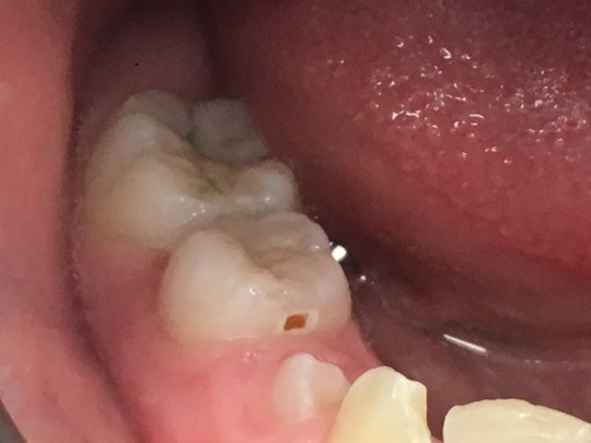
Fig. 5. Caries
Note
Baby bottle tooth decay is found in very young children who regularly have been given a nursing bottle, who have experienced prolonged breasts feeding, used pacifier dipped in a sweet agent. Maxillary anterior teeth and primary molars are affected. The source of the problem can be detected and preventive procedures started with parenteral education.
1.2.2
Root surface cavity
Root surface cavity is a soft lesion of cementum and dentin which involves bacterial infection and invasion. Gingival recession is necessary for root caries. Caries does not form in the root surface while periodontal fibres are still attached.
+
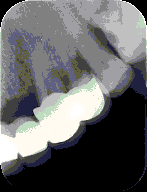
Fig. 6. Root caries
Formation of a cavity
Gingival recession exposes the cemental surface. Mutans streptococci and lactobacilli are organisms associated with root caries. Cementum is very thin and is soon destroyed. Dentin is invaded. It increases and extend completely around the tooth with undermining of the enamel. They are described as soft, leathery, or hard lesions. Active lesions are soft or leathery, inactive are hard.
Risk factors of root caries
The factors involved daily inadequate personal hygiene with bacterial plaque accumulation, frequent use of cariogenic foods, insufficient intake of fluoride, periodontal infection. The others can be using medications with side effects, radiation to head, many restorations, overhanging margins, and prosthetic devices.
1.2.3
Treatment of cavities
Regular check-ups can identify cavities before they cause painful symptoms. Treatment of cavities depends on how severe they are.
Options can be:
- Fluoride treatment. Low concentrations of fluoride should be applied frequently during the early phase. It can provide sources for uptake by the demineralized zone. This place takes fluoride from dentifrice, mouthwashes, fluoridated drinking water, or special gel or foam which is placed in a small tray that fits over patient´s teeth.
- Ozone therapy. Ozone is toxic to certain bacteria and delivering ozone into a carious lesion might reduce the number of cariogenic bacteria. It could arrest the progress of the lesion and may, in the presence of fluoride, allow remineralisation.
- Restorations (fillings) are the main treatment possibility when decay has progressed beyond the earliest stage. Fillings are made of various materials. Tooth-coloured composite resins, porcelain or dental amalgam, glass ionomer cement, etc.
- Crown. If the tooth is weak, or decay is extensive, the crown is used. It is a custom-fitted covering that replaces patient´s natural crown. It may be made of high strength porcelain, resin, porcelain fused to metal, or gold.
- Root canals. When decay reaches the pulp, the root canal is needed. The diseased pulp is removed, medication can be used in root canal to clear infection, and finally the pulp is replaced with a filling.
- Tooth extraction. Sometimes the tooth is so decayed that it can´t be restored. Pulled tooth can leave a gap. To avoid shifting of other teeth the patient can get a bridge, or dental implant to replace missing tooth.
Summary
The sooner the patient seek care, the better the chances are for reversing the earliest stages of tooth decay and preventing its progression.
1.2.4
Noncarious dental lesions
Erosion
Erosion is the loss of tooth substance by a chemical process that does not involve known bacterial action. It is smooth, shallow, hard, shiny place. It is usually found on facial or lingual surfaces. It depends on the cause, which can be chronic vomiting (because of pregnancy, or eating disorder). The lingual surfaces are affected. Extrinsic factors affect usually facial surfaces (worker´s teeth can be exposed to atmospheric acids, or carbonated beverages or lemon juice used frequently). It may occur in combination with dental caries, or calculus.
+
Fig. 7. Erosion in a nine-year-old patient with oesophageal reflux
Abrasion
Abrasion is the mechanical wearing away of tooth substance by forces other than mastication. The lesions originate from a mechanical abrasive activity. The appearance can be V shaped or edge shaped with hard, smooth, shiny surface and clearly defined margins. Dental caries may occur in this area as a secondary lesion. The most common cause is an abrasive dentifrice applied with wrong brushing technique. Other possibility can be occupational cause (pins held between teeth by dressmakers), or pipe smokers hold pipe in the same place over many years.
Attrition
Attrition is a physiological wearing away of the biting surfaces due to tooth-to-tooth contact or a long-term contact of the teeth with some coarse materials. It is caused by tooth clenching, working in the environment where coarse dust constantly gets into the mouth, tooth grinding.
Demineralization
White spots or dental caries have been common findings after orthodontic treatment. Bacterial plaque retention by the appliances and the resin, along with difficulty of plaque removal by the patient, contribute to demineralization and dental caries. The configurations of the appliances make plaque control efforts by the patient extremely difficult. Plaque collects on the brackets and some resins even when the patient´s hygiene is good. The surface of composite resin, which can be left around the brackets, is difficult to make smooth. That´s why plaque forms here. After de-bonding, the use of retainer is a cause of another retention of bacterial plaque.
+
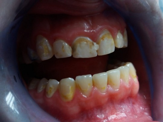
Fig. 8. Demineralization after braces
DH should teach the patient how to apply the toothbrush and auxiliary devices to remove plaque from the bracket, the arch wire, and the teeth. How, when, and why to use fluoride rinse, toothpaste, and brush-on fluoride gel.

Video 1. Brushing teeth with braces (the user will be directed to an external page)
©
For licensing reasons, this interactive object cannot be directly incorporated into the material. Click HERE to see the object.
Interactive object 2. Type the answer in English (the user will be directed to an external page)